
Reflex arc Medical school inspiration, Medical student study, Medical school studying
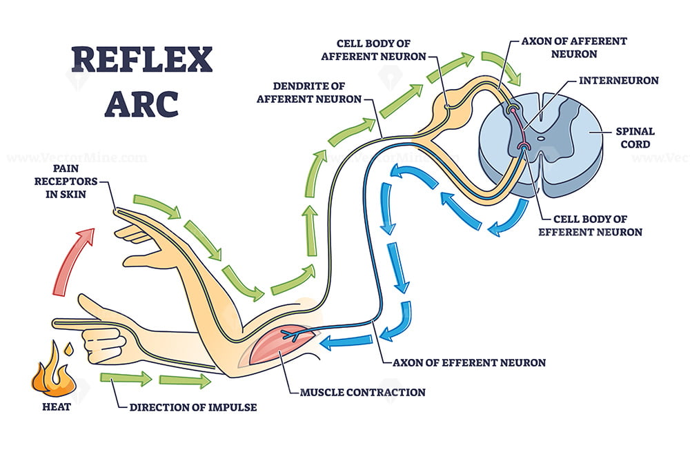
A reflex arc is a neural pathway that controls a reflex. In vertebrates, most sensory neurons do not pass directly into the brain, but synapse in the spinal cord. This allows for faster reflex actions to occur by activating spinal motor neurons without the delay of routing signals through the brain.
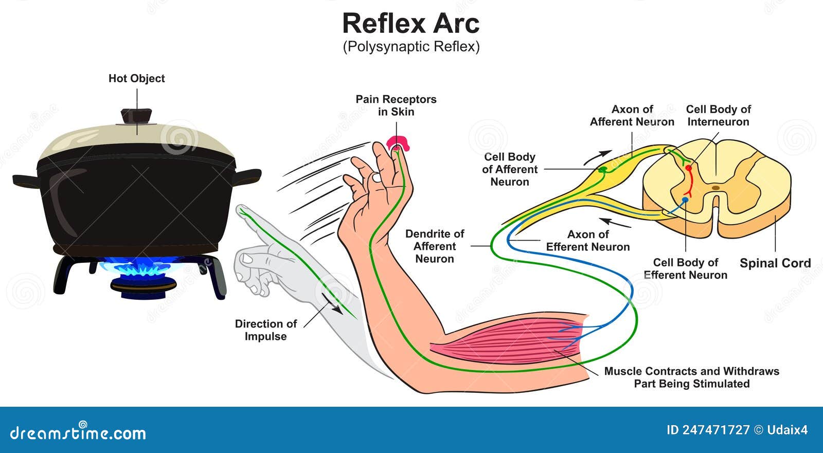
Reflex Action and Reflex Arc What Happens When You Accidentally Touch a Hot Pot Owlcation
A reflex arc occurs when the body responds automatically to an outside stimulation. When someone touches a hot surface, the body responds utilizing a reflex arc to remove the body from the high.
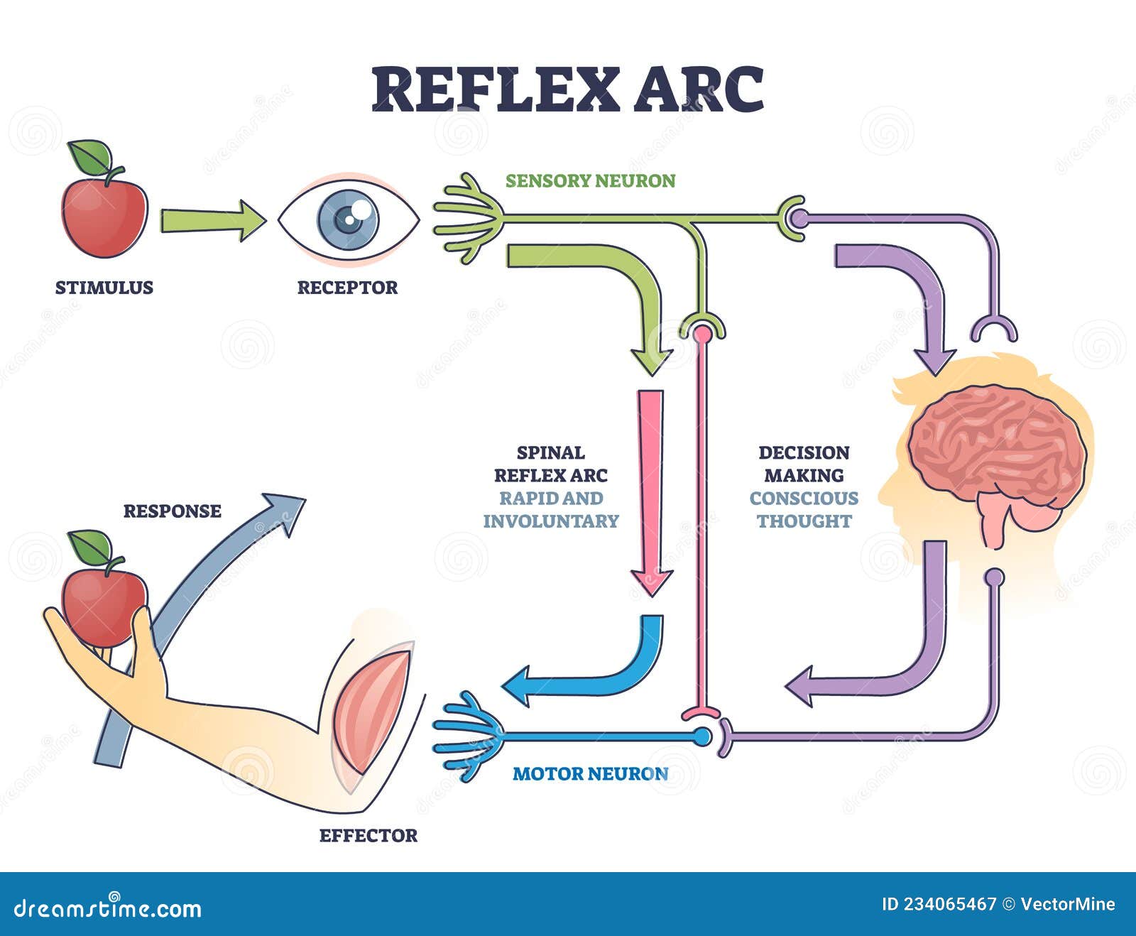
Reflex ARC Sensory Neuron Pathway from Stimulus To Response Outline Diagram Stock Vector
The reflex arc is the pathway that a signal follows from stimulus to response during a reflex action. The typical reflex arc of a simple reflex has seven components, which are shown in Figure 2. Figure 2 : A flow chart showing the 7 components of a reflex arc, from the stimulus to the response.

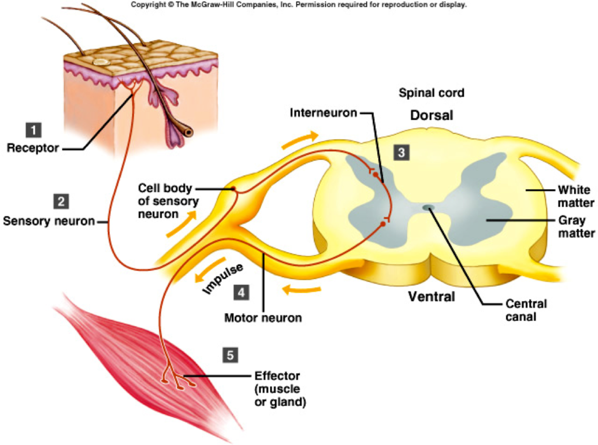
How to Draw Reflex Arc Reflex Arc Diagram Structure of Reflex Arc in Very Simple Way YouTube
A reflex arc refers to the neural pathway that a nerve impulse follows. The reflex arc typically consists of five components: A receptor, and independent sensory cell, or an ending of a sensory neuron, reacts to a stimulus (e.g., a stretch receptor).. Circuit diagram for recording electromyograms from the calf muscles. Make sure the ankle.

20182019 Marieb Reflex Arc Diagram Quizlet
The primary components of the reflex arc are the sensory neurons (or receptors) that receive stimulation and in turn connect to other nerve cells that activate muscle cells (or effectors), which perform the reflex action.

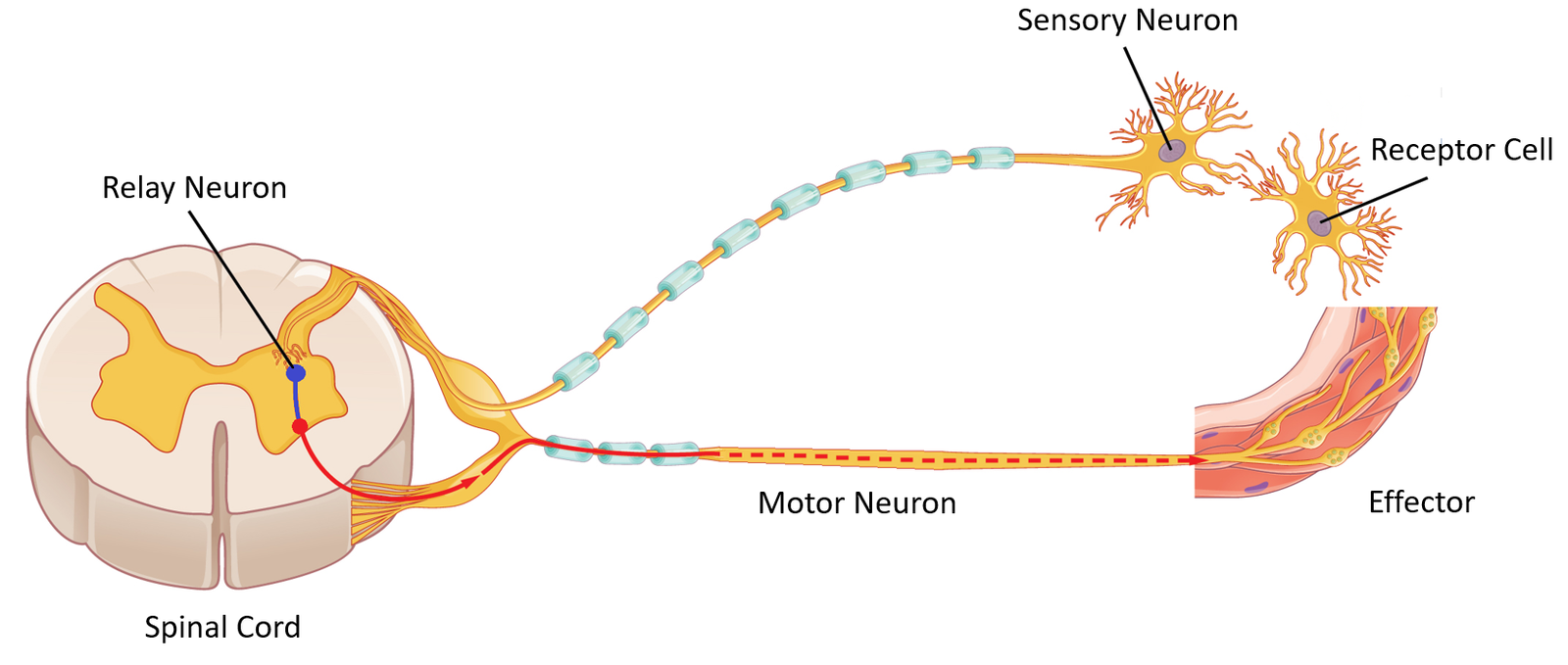
Schematic representation of a spinal reflex arc. A pin in the skin... Download Scientific Diagram
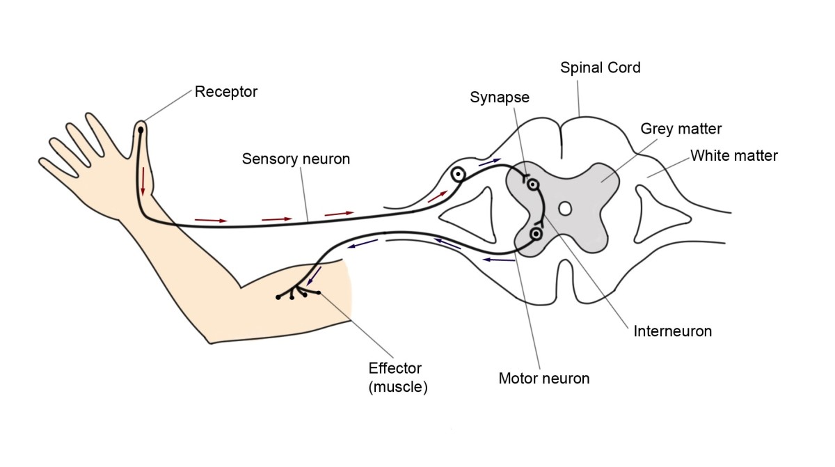
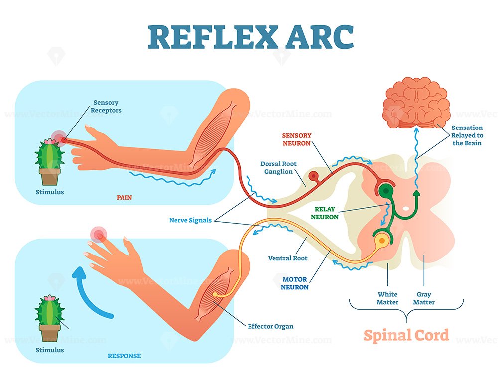
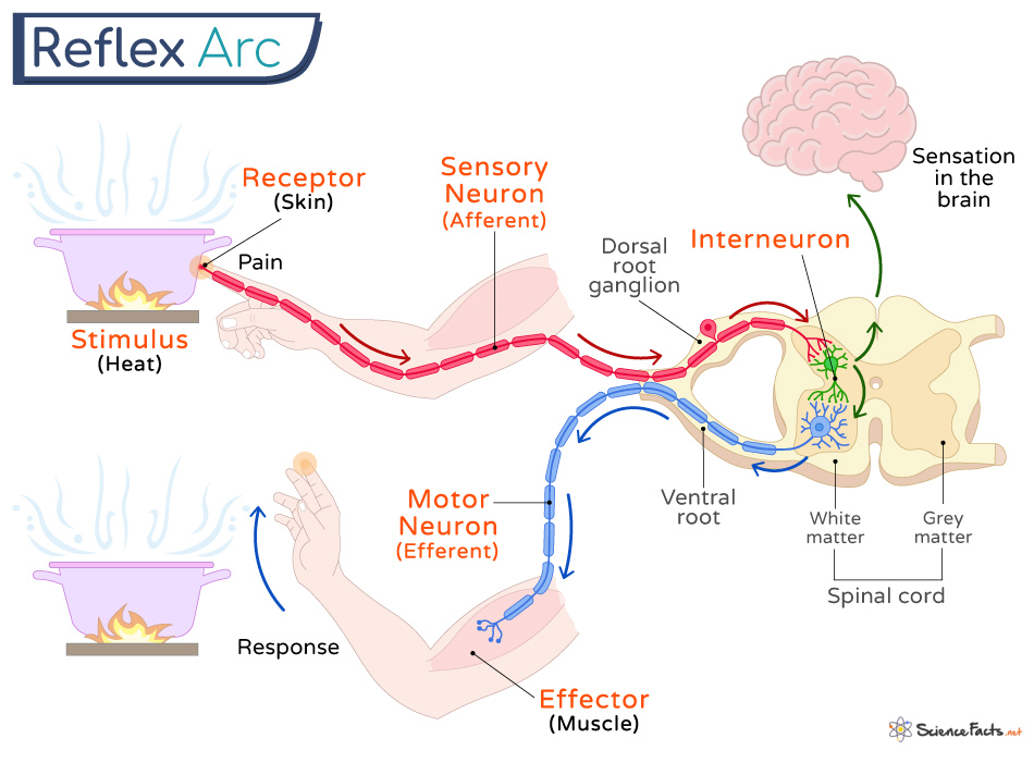
• Drawing and labelling a diagram of a reflex arc for a pain withdrawal reflex In a pain withdrawal reflex arc: A pain stimulus is detected by a receptor (nocireceptor) and a nerve impulse is initiated in a sensory neutron The sensory neuron enters the spinal cord via the dorsal root and synapses with a relay neuron in the grey matter

2. The mammalian stretch reflex arc. Download Scientific Diagram
By definition, a reflex is an involuntary, stereotypical response of the effector tissue from the stimulation of receptors. These reflexes are executed by the successive activation of a certain number of neurons that are mutually connected.
All About The Spinal Cord and Its Importance HubPages
Labelled Diagram Of A Reflex arc Reflex Arc Diagram This labelled diagram of a reflex arc indicates the neural pathway controlling a reflex. It clearly indicates the route adapted when a stimulus occurs and how the reaction takes place.
Reflex Arc Key Stage Wiki
There are three main types of neurone in a reflex arc: sensory, relay and motor Sensory neurones carry impulses from sense organs to the CNS (brain or spinal cord) Relay neurones are found inside the CNS and connect sensory and motor neurones Motor neurones carry impulses fr om the CNS to effectors (muscles or glands)
Reflex arc explanation with pain signals and receptor impulse outline diagram VectorMine
A reflex action is an automatic (involuntary) and rapid response to a stimulus, which minimises any damage to the body from potentially harmful conditions, such as touching something hot..
Reflex Arc Polysynaptic Infographic Diagram Stock Vector Illustration of infographic, body
Reflex Arc A signal travels from the organ and initiates a response to the organ that reacts to the signal. This response causes various parts of the reflex arc to work in order. A simple reflex arc has the following parts: 1. Stimulus It is any change in the environment (internal or external) detected by a receptor.
😍 Explain a reflex arc. How Does Reflex Arc Work?. 20190129
Below is a diagram of a reflex arc: a. Label each cell as an interneuron, motor neuron or sensory neuron. b. Using arrows, indicate the direction of nerve impulse through each neuron. c. Label the receptor end of the sensory neuron, the dendrite and axon. d. Label the effector (muscle/organ) of the motor neuron, the dendrite and the axon.
Spinal Reflex Arc anatomical scheme, vector illustration VectorMine
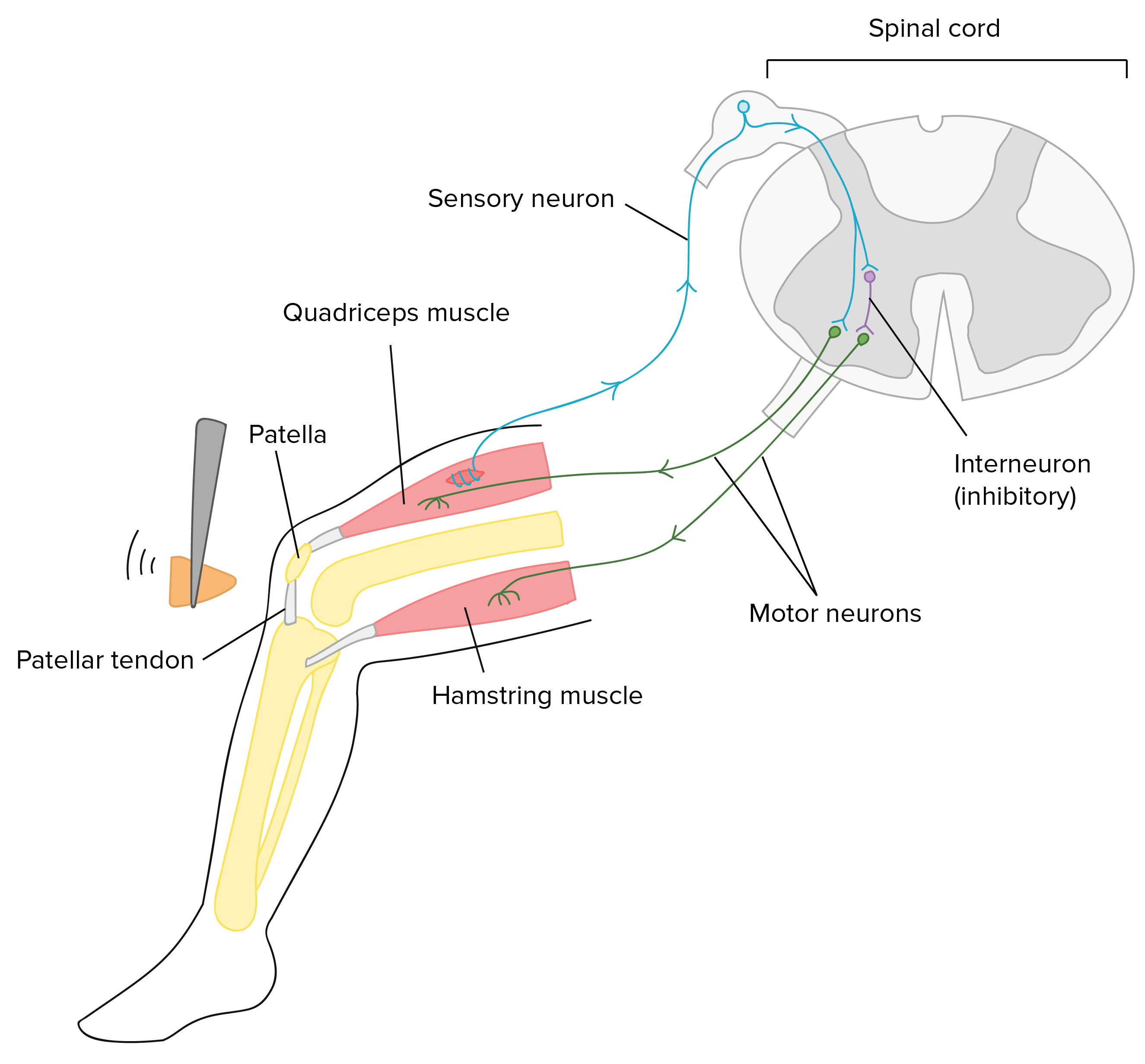
Key Points. Reflexes, or reflex actions, are involuntary, almost instantaneous movements in response to a specific stimulus. Reflex arcs that contain only two neurons, a sensory and a motor neuron, are considered monosynaptic. Examples of monosynaptic reflex arcs in humans include the patellar reflex and the Achilles reflex.
Reflex Arc Definition, Steps, Components, and Diagram
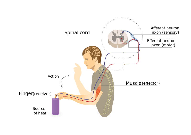
fig 1 - Diagram demonstrating the components of a reflex arc and the reflex response to a heat source. The Monosynaptic Stretch Reflex A monosynaptic reflex, such as the knee jerk reflex, is a simple reflex involving only one synapse between the sensory and motor neurone.
5 Elements of a Reflex Arc
reflex arc . For example, a simple reflex arc happens if we accidentally touch something hot. Receptor in the skin detects a stimulus (the change in temperature). Sensory neurone sends.

Think Tank Centre The Reflex Arc
How does the human body work? - Class 11 Course: How does the human body work? - Class 11 > Unit 6 Lesson 3: Human nervous system Brain: Parts & functions (Fore, mid & hind) Overview of the functions of the cerebral cortex Thalamus, hypothalamus, and limbic system Reflex action (& reflex arc) Functions of brain Forebrain and reflex action Science >
