Arteries vs Veins Structure, Function & Blood Flow
1/7 Synonyms: Dorsal thalamus, Thalamencephalon , show more. Cross-sections are two-dimensional, axial views of gross anatomical structures seen in transverse planes. They are obtained by taking imaginary slices perpendicular to the main axis of organs, vessels, nerves, bones, soft tissue, or even the entire human body.
:max_bytes(150000):strip_icc()/the-structure-of-the-vein-wall-87395965-58963cc63df78caebc05eee0.jpg)
Superior and Inferior Venae Cavae
The short-axis cross-sectional areas of the subclavian vein at the mid-clavicular line, the subclavian vein in the supraclavicular fossa, and the internal jugular vein at the level of the thyroid cartilage were calculated. Results

Similarities and Differences Between Arteries and Veins Facty Health
Browse 2,975 authentic vein cross section stock photos, high-res images, and pictures, or explore additional artery cross section or blood vessel stock images to find the right photo at the right size and resolution for your project. Browse Getty Images' premium collection of high-quality, authentic Vein Cross Section stock photos, royalty-free.

Artery and Vein Cross Section Diagram Quizlet
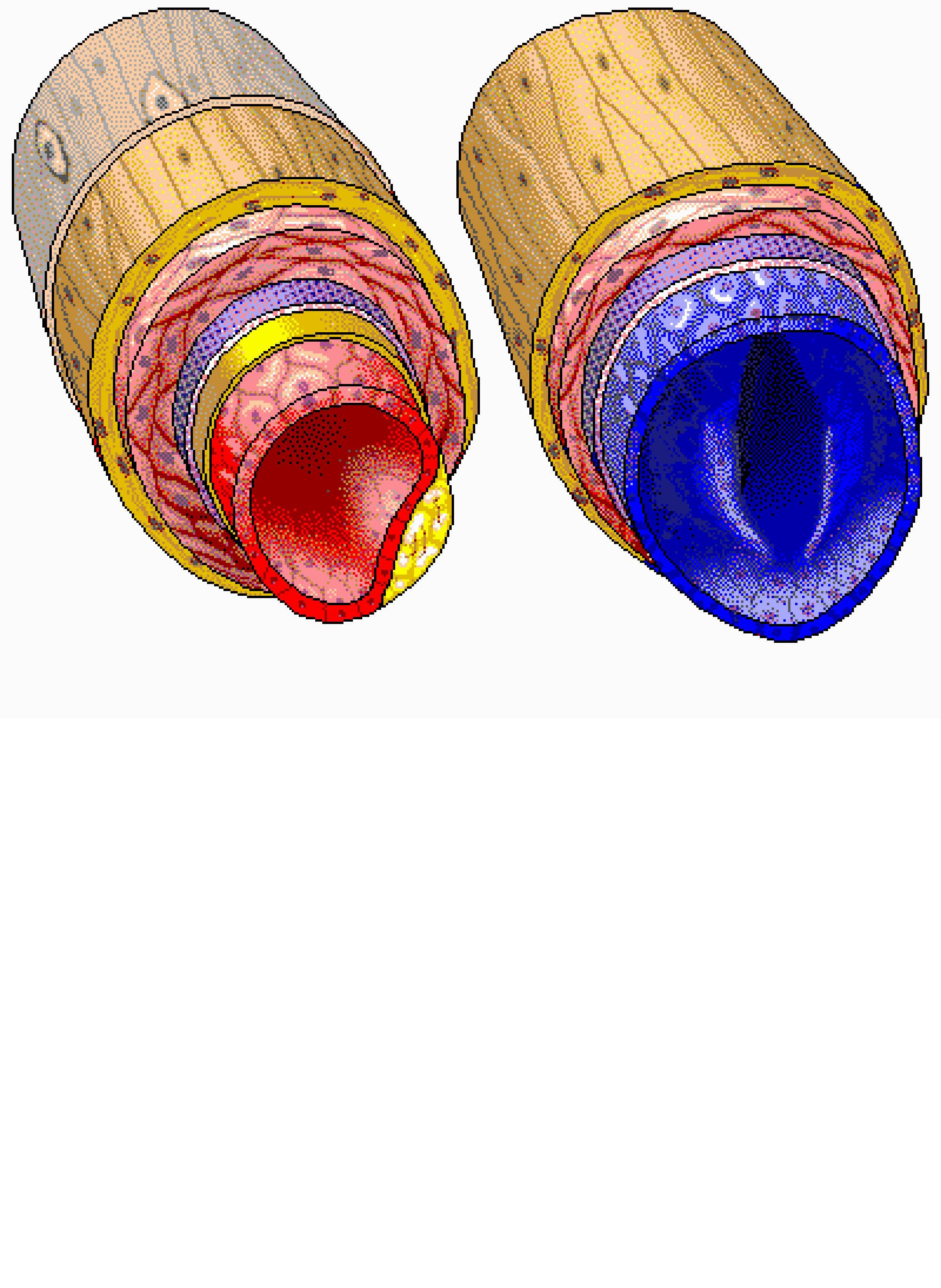
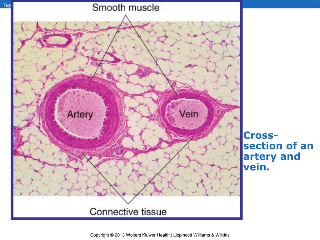
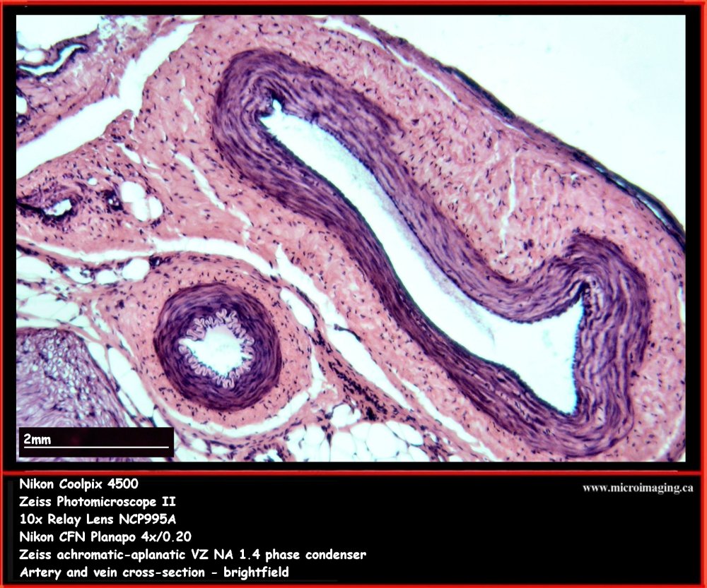
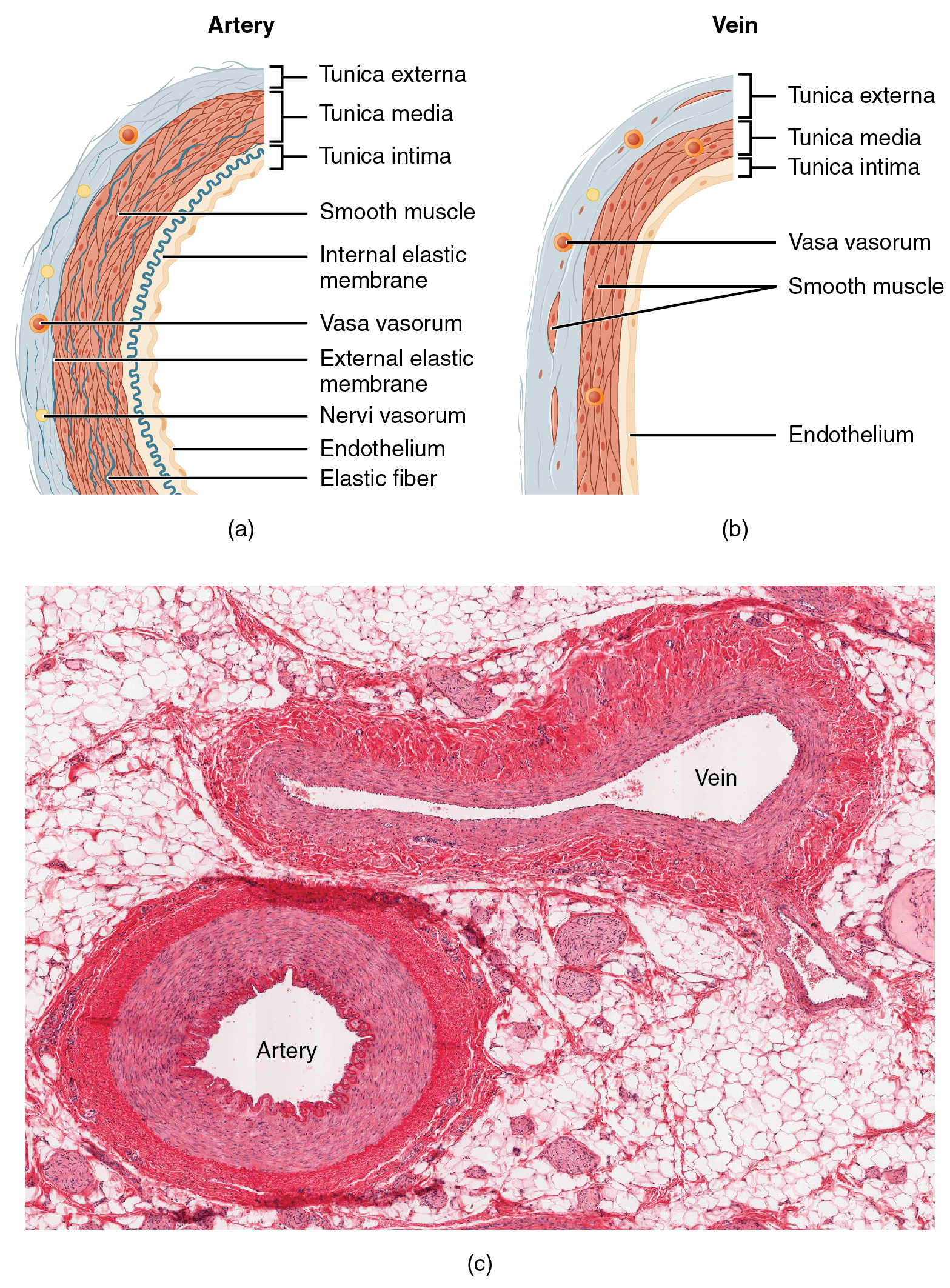
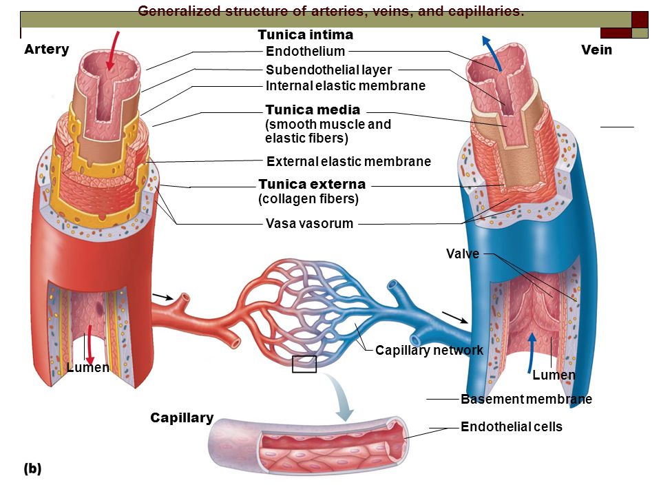
Together, their thicker walls and smaller diameters give arterial lumens a more rounded appearance in cross section than the lumens of veins. Figure 20.3 Structure of Blood Vessels (a) Arteries and (b) veins share the same general features, but the walls of arteries are much thicker because of the higher pressure of the blood that flows through.

Micrograph illustrating a cross section of a medium size muscular artery, its vein
Together, their thicker walls and smaller diameters give arterial lumens a more rounded appearance in cross section than the lumens of veins.. Figure \(\PageIndex{9}\): Varicose Veins. Varicose veins are commonly found in the superficial veins of the lower limbs. Defective valves cause localized pooling of blood in the veins that can lead to.

Arteries And Veins Cross Section
The azygos venous system is located on either side of the vertebral column and drains the viscera within the mediastinum, as well as the back and thoracoabdominal walls. This system consists of the azygos vein and its two main tributaries: the hemiazygos vein and the accessory hemiazygos vein.

What are Blood Vessels? Types, Structure, & Functions hubpages
This article is a comprehensive CT-based imaging review of the pulmonary veins, including their embryology, anatomy (typical and anomalous), surgical implications, pulmonary vein thrombosis, pulmonary vein stenosis, pulmonary vein pseudostenosis, and the relationship of tumors to the pulmonary veins. Online supplemental material is available.

artery/vein cross section practical Diagram Quizlet
PURPOSE: To retrospectively establish normal values for pulmonary vein diameter, cross-sectional area, and shape depicted at computed tomography (CT). MATERIALS AND METHODS: Institutional review board waived patient consent requirement and approved the study. Thin-section contrast material-enhanced spiral chest CT scans in 104 patients, 68 women and 36 men (age range, 19-86 years; mean, 49.
PPT Chapter 14 Blood Vessels and Blood Circulation PowerPoint Presentation ID5387380
Veins are composed of xylem and phloem cells embedded in parenchyma, sometimes sclerenchyma, and surrounded by bundle sheath cells. The vein xylem transports water from the petiole throughout the lamina mesophyll, and the phloem transports sugars out of the leaf to the rest of the plant.

Blood Vessel Structure and Function Boundless Anatomy and Physiology
Find an appropriate vein by scanning the arm in the transverse orientation, which provides a cross-sectional view of the anatomy and allows simultaneous visualization of veins, arteries, and other.

LM of a crosssection through an artery and vein Stock Image P206/0116 Science Photo Library
Lining the core of each is a thin layer of endothelium, and covering each is a sheath of connective tissue, but an artery has thick intermediate layers of elastic and muscular fiber while in the vein, these are much thinner and less developed. With the exception of pulmonary and umbilical veins and arteries, arteries carry oxygenated blood from.
Artery & Vein, cross section
Blood vessel histology Author: Lorenzo Crumbie MBBS, BSc • Reviewer: Dimitrios Mytilinaios MD, PhD Last reviewed: October 30, 2023 Reading time: 17 minutes It would be impossible to get blood to the predestined locations without the vascular pathways. Blood vessels form the extensive networks by which blood leaves the heart to supply tissue. . Additionally, other blood vessels return from.
20.1 Structure and Function of Blood Vessels Douglas College Human Anatomy and Physiology I
Figure 40.10.1 40.10. 1: Blood vessel layers: Arteries and veins consist of three layers: an outer tunica externa, a middle tunica media, and an inner tunica intima. Capillaries consist of a single layer of epithelial cells, the endothelium tunic (tunica intima). Veins and arteries both have two further tunics that surround the endothelium: the.

Structure and Function of Blood Vessels Anatomy and Physiology II
The short-axis (cross-sectional, transverse) ultrasound view is easy to obtain and is the better view for identifying veins and arteries and their orientation to each other.. Cannulate a central vein at a site of optimal short-axis imaging (ie, large-diameter cross section of the vein, with no overlying artery). Attach the cardiac monitor to.

Vein Crosssection. Lm Photograph by Science Stock Photography Fine Art America
The presence of the costal vein was not observed on the cross-section during the present study. Our results are consistent with those produced for Orthezia urticae by Franielczyk-Pietyra et al. . The costal vein has not been recognized in wings of scale insects so far, e.g., [16,27,29,30].
Blood Vessels Alisa Houghton
The right hepatic vein is a single dominant vein in ~70% (range 60-78%) of individuals. There may be an early bifurcation, early trifurcation or even multiple right hepatic veins entering the IVC. Hence this may make it difficult to accurately deduce segmental anatomy of the liver. The commonest anatomical variant of the hepatic veins is an.
