Multiple Myeloma Stepwards
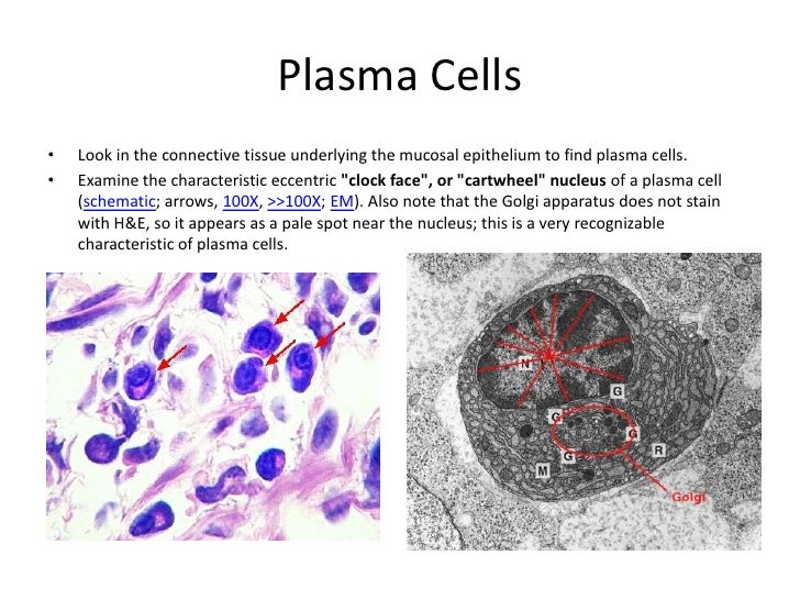
The plasma cell clock-face or cart-wheel nuclear pattern as seen in 2D sections/projections from almost any angle can be explained by the multiradial arrangement of peripherally placed clump units.

Lin’s Lab Unravels the Pathway of Plasma Cells
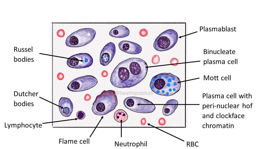
Mott cells are plasma cells that have spherical inclusions packed in their cytoplasm. The term 'Mott cell' is named after a surgeon, F. W. Mott, who identified these cells in the brains of monkeys with trypanosomiasis (1901). He termed it morular cell (from the Latin morus, mulberry) and recognized these cells to be plasma cells and.

Plasmablastic lymphoma cells with a plasmacytoid appearence with a... Download Scientific Diagram
Ovoid intermediate-small cell size ~ 12 micrometers: Cells slightly larger than red blood cells and neutrophils. Eccentric nucleus. Nucleus usu. hugs the cell membrane. "Clock-face" chromatin pattern. Small dots symmetrically rim the nuclear membrane - like the numbers on a clock. Abundant cytoplasm. Nucleus-to-cytoplasm ratio ~1:2.
Electron Micrograph
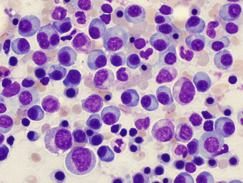
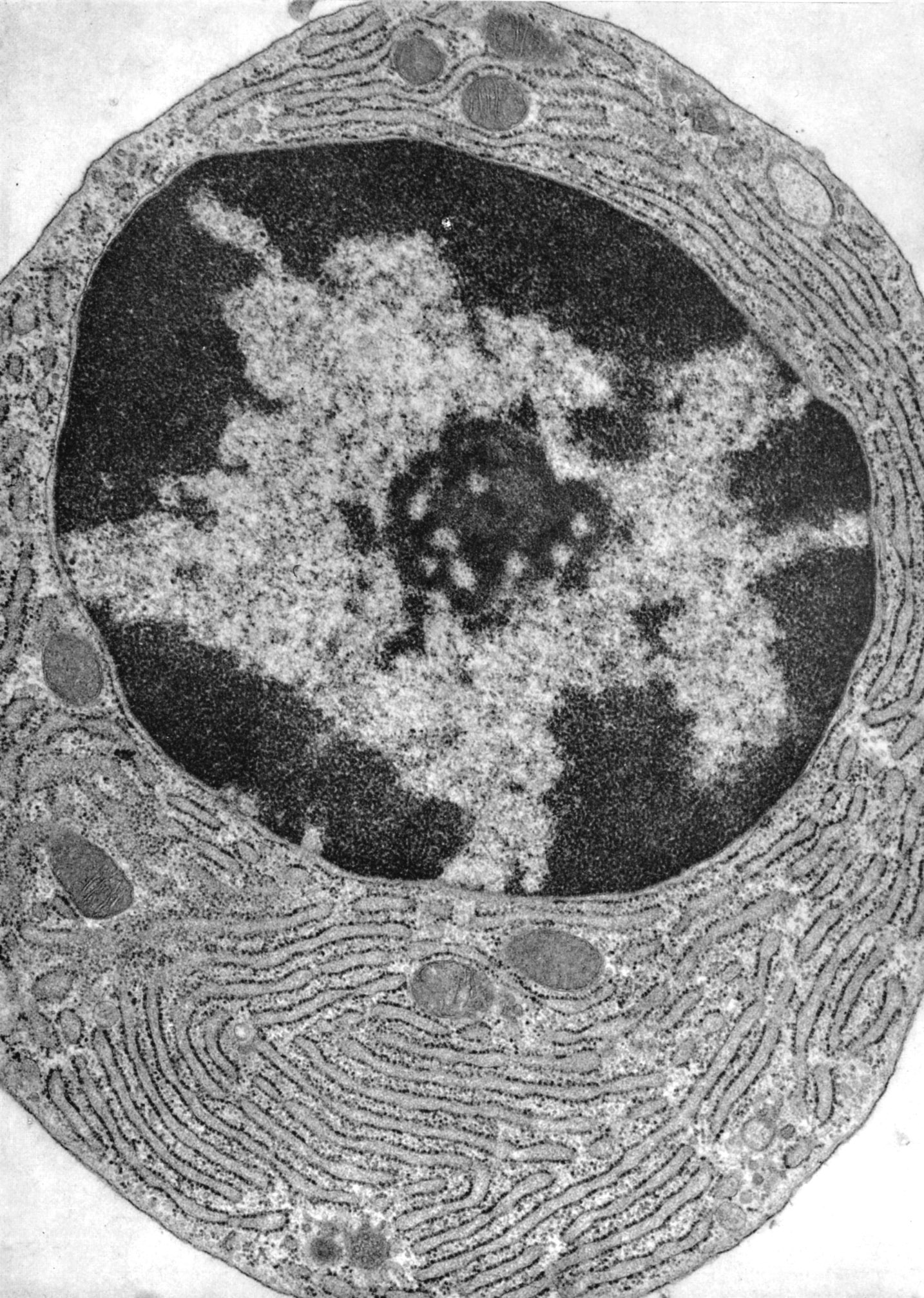
Plasma cells are 14 to 20 microns in diameter and are characterized by a strongly basophilic cytoplasm. Electron microscopy reveals an eccentrically-located nucleus that has a well-developed nucleolus and variable amounts of condensed chromatin. Chromatin associated with the nuclear membrane gives the nuclei the appearance of a clock-face or.

Sorting strategy of plasma cells representative flow cytometry plots... Download Scientific
Clock-Face Chromatin Pattern: Within the nucleus, the chromatin forms clumps or patches, resembling a clock-face arrangement. This pattern is a characteristic feature of plasma cells and distinguishes them from other immune cell types. Localization of Plasma Cells: Plasma cells are found in various anatomical locations within the body, including:
Samantha blum histo study guide 2
An accumulation of plasma cells demonstrates two diagnostic features that help identify plasma cells. Heterochromatin frequently clumps around the periphery of the nucleus, forming a "clock face" appearance. Additionally, a pale-staining region in the cytoplasm adjacent to the nucleus indicates the position of the large Golgi apparatus.
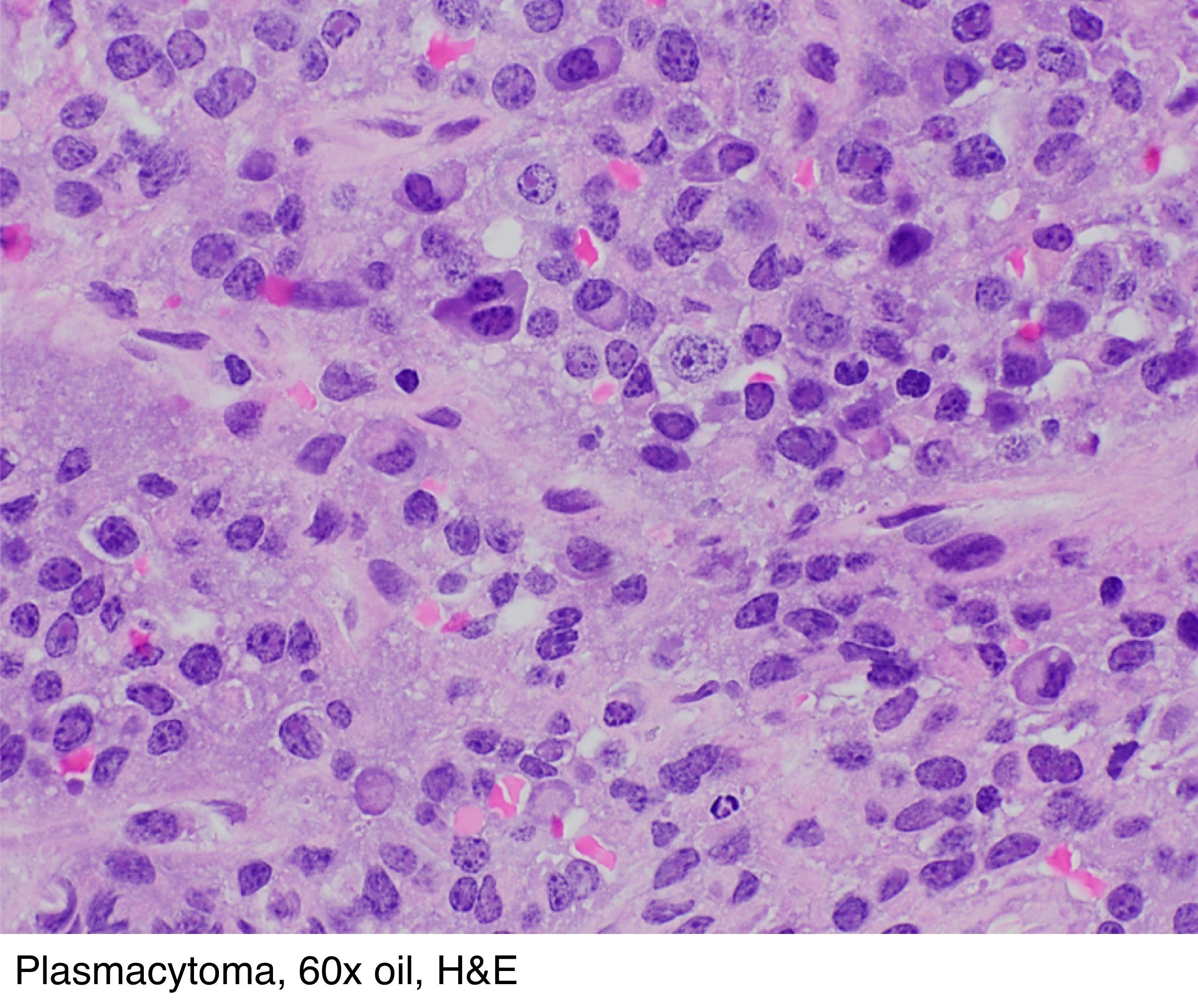
Pathology Outlines Plasmacytoma
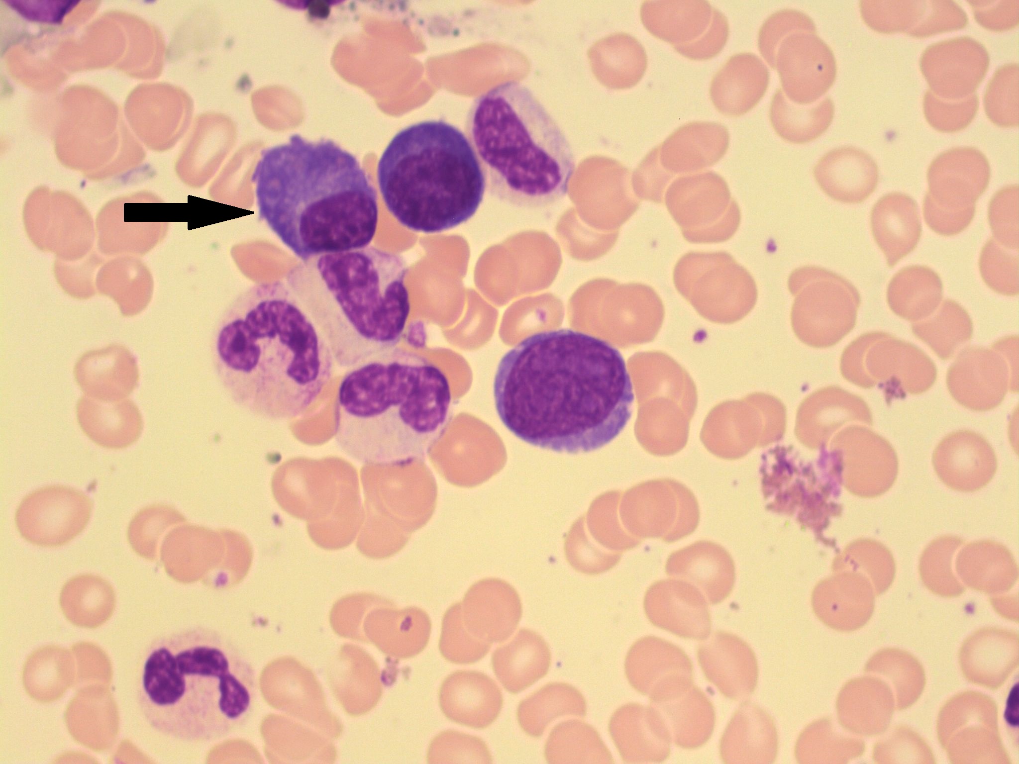
Plasma cells - 1. #00004107. Author: Peter Maslak. Category: Lymphoma: Mature B-cell and Plasma cell Neoplasms > Plasma Cell Neoplasm. Published Date: 11/01/2009. Plasma cells (arrows) have eccentric nuclei characterized by a "clock face" appearance to the chromatin. The cytoplasm may range from basophilic to blue-gray and can contain vacuoles.

Figure 14 from Plasma cell morphology in multiple myeloma and related disorders. Semantic Scholar
Plasma cells have distinctive features that are clearly seen in this electron micrograph: a prominent Golgi; well developed rough endoplasmic reticulum; and a nucleus with large clumps of heterochromatin at the margin of the nucleus (clock-face nucleus). Compare these features with the high magnification light microscopic inset. Plasma cells.
Plasma cells 1.
Download scientific diagram | Plasma cells are identified by their eccentric, clock-face nucleus and pale perinuclear cytoplasmic crescent. Staining by hematoxylin-eosin stain (magnification 400×.

Cell structure Wall Clock by Admin_CP66866535
However, the nuclei of these plasma cells are eccentric with a morphology typical of plasma cells (coarse chromatin, arranged in a clock face pattern). Immunophenotyping of these cases is very helpful as plasma cells in this variant are positive for CD138, in contrast to histiocytes in crystal storing histiocytosis which are positive for.
The plasma cell clockface or cartwheel nuclear pattern as seen in 2D... Download Scientific
Structure Plasma cells with Dutcher and Russell bodies (H&E stain, 100×, oil). Plasma cells are large lymphocytes with abundant cytoplasm and a characteristic appearance on light microscopy.They have basophilic cytoplasm and an eccentric nucleus with heterochromatin in a characteristic cartwheel or clock face arrangement. Their cytoplasm also contains a pale zone that on electron microscopy.

2 Face collection. Plasma Pulse. WatchMaker Watch Faces
The nucleus of the plasma cell is spherical and usually eccentrically positioned. It contains large clumps of peripheral heterochromatin interspersed with clear areas of euchromatin, giving it a characteristic cartwheel or an analog clock face appearance.
Plasma Cells A Laboratory Guide to Clinical Hematology
The first round of anti-CD20 depletion was performed at 1.5 years after vaccination and when tetanus-specific memory B cell frequencies were determined at 1.7 years (2.5 months after depletion.
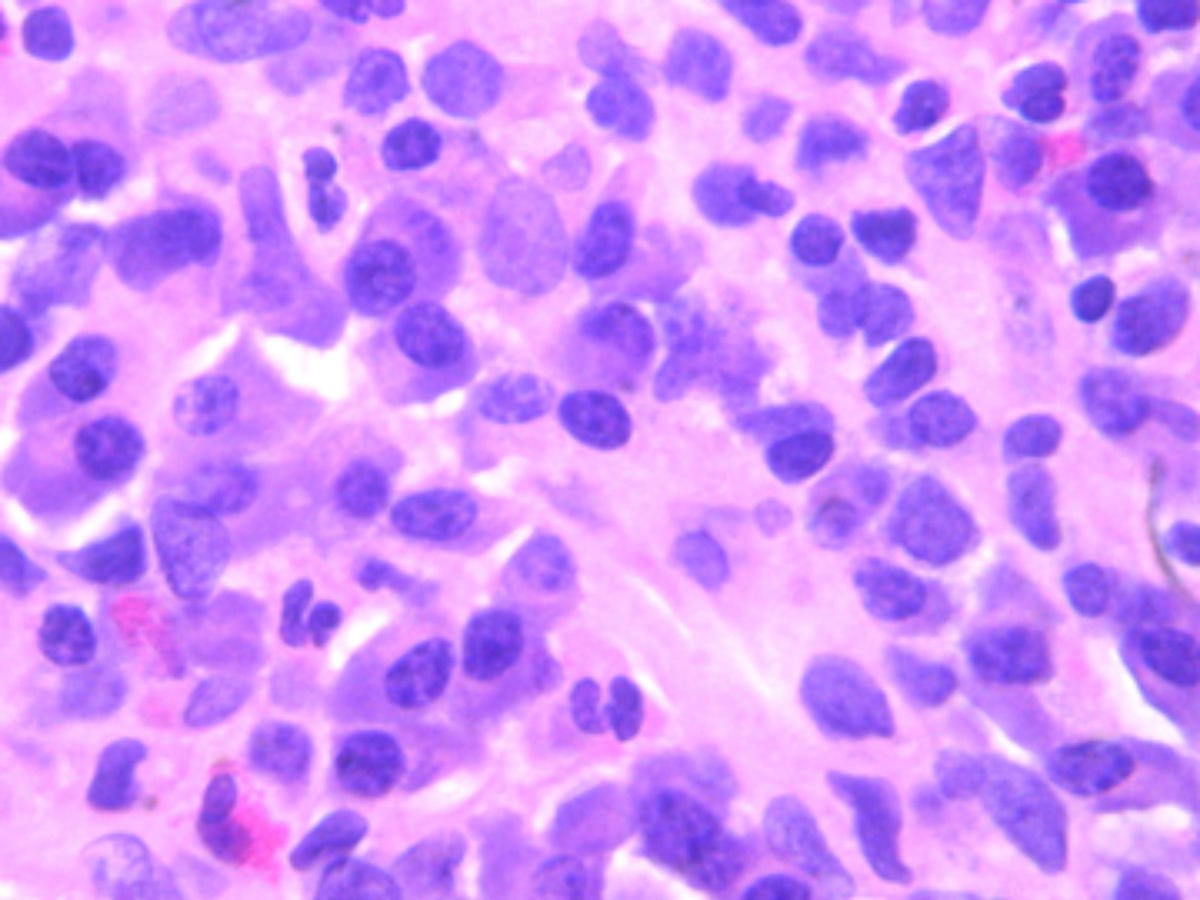
Pathology of Multiple Myeloma Pathology Made Simple
Definition / general. Usually less than 1% of marrow cells; rare in infants. Often perivascular and in particle crush specimens. Indeterminate lifespan ranging from days to months. Produces and secretes antibodies. Plasmablast: precursor to plasma cell, produces more antibodies than B cells but less than mature plasma cells.
An unusual presentation of Castleman's Diseasea case report BMC Infectious Diseases Full Text
Multiple Myeloma is neoplastic proliferation of plasma cells that commonly results in multiple skeletal lesions, hypercalcemia, renal insufficiency, and anemia. Patients typically present at ages > 40 with localized bone pain or a pathologic fracture. Diagnosis is made with a bone marrow biopsy showing monoclonal plasma cells ≥10%.

Figure 17 from Plasma cell morphology in multiple myeloma and related disorders. Semantic Scholar
Plasma cells vary in size from 14 to 20 micrometers. They are round-to-ovoid cells containing abundant deep blue cytoplasm with a pale perinuclear area corresponding to the Golgi apparatus. They have a round, eccentrically placed nucleus with coarse chromatin arranged in a clock face (art wheel) pattern. Most plasma cells are uninucleate; few.
