Solved Chapter 1, Problem 03 A Visual Analogy Guide to Human Anatomy
Tissue Membranes. The two broad categories of tissue membranes in the body are (1) connective tissue membranes, which include synovial membranes, and (2) epithelial membranes, which include mucous membranes, serous membranes, and the cutaneous membrane—in other words, the skin. From Betts, et al., 2013. Licensed under CC BY 4.0.
Body Cavity Diagram ClipArt Best
Body cavities diagram from an animal. You already got a different diagram of the body cavity from an animal. Here, I will show you again some of the cavities in one diagram. But, if you need more updated diagrams on the body cavity, you may join anatomy learner on social media..

Dorsal body cavity Wikipedia
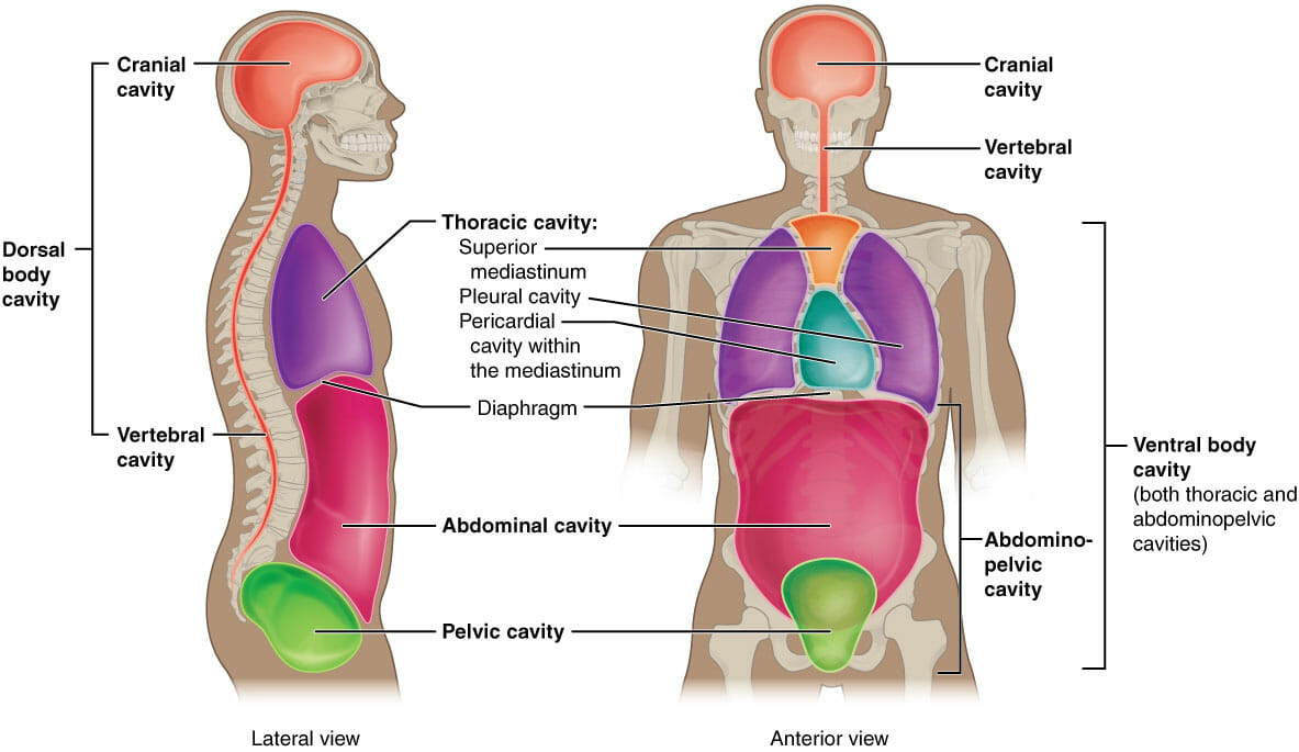
A body cavity is any space or compartment, or potential space, in an animal body. Cavities accommodate organs and other structures; cavities as potential spaces contain fluid. The two largest human body cavities are the ventral body cavity, and the dorsal body cavity. In the dorsal body cavity the brain and spinal cord are located.

What Are the Different Types of Human Body Cavities?
Figure 33.6.1 33.6. 1: Body planes: Shown are the planes of a quadruped goat and a bipedal human. The midsagittal plane divides the body exactly in half into right and left portions. The frontal plane divides the front and back, while the transverse plane divides the body into upper and lower portions. Vertebrate animals have a number of.
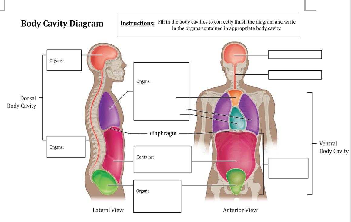
Solved Body Cavity Diagram Instructions Fill in the body
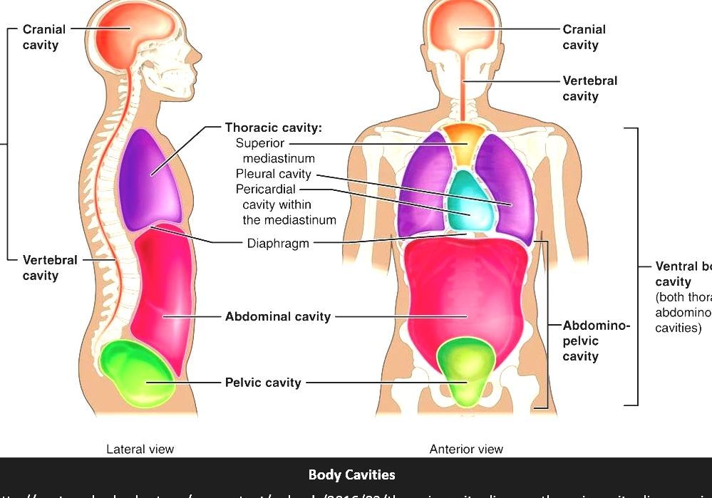
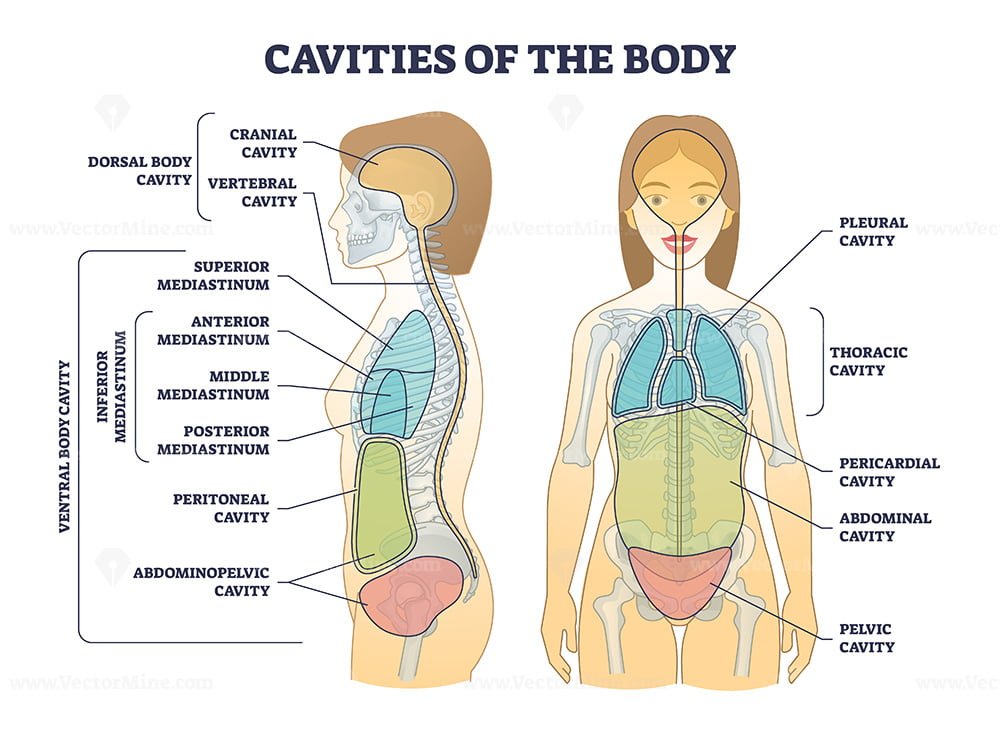
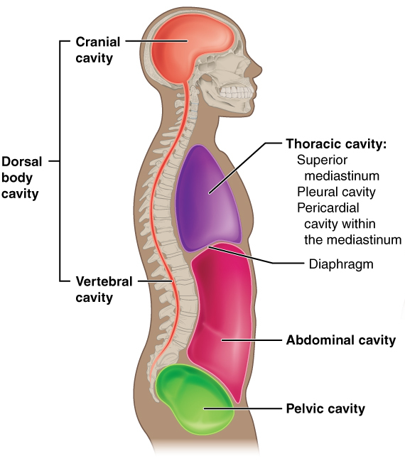
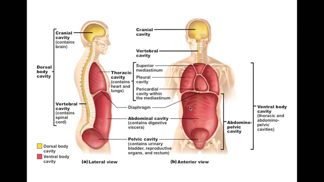
A. body cavity. is a fluid-filled space inside the body that holds and protects internal organs. Human body cavities are separated by membranes and other structures. The two largest human body cavities are the ventral cavity and dorsal cavity. These two body cavities are subdivided into smaller body cavities.
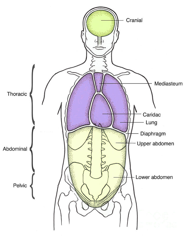
Illustration Of Anterior Body Cavities Photograph by Science Source
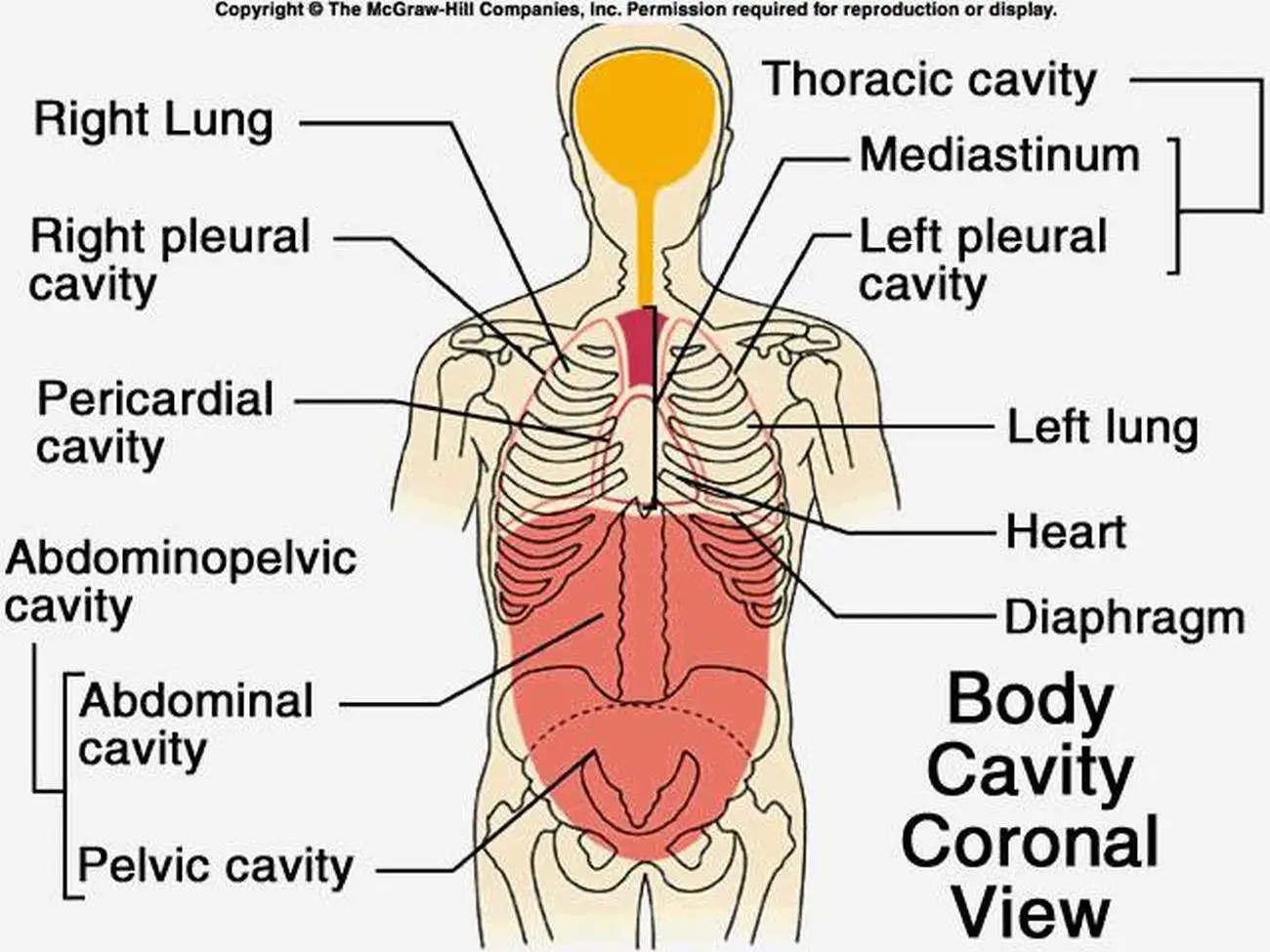
The thoracic cavity is the anterior ventral body cavity found within the rib cage in the torso. It houses the primary organs of the cardiovascular and respiratory systems, such as the heart and lungs, but also includes organs from other systems, such as the esophagus and the thymus gland. The thoracic cavity is lined by two types of mesothelium.

Body Cavities Chest Cavity Anatomy and Body Cavities Anatomy
Discover the body's cavities and organs found in the ventral cavity, where it is located, and see a ventral body cavity diagram. Updated: 11/21/2023 Table of Contents. What is the Ventral Cavity?.
Body Cavities and Organs Biology Dictionary
The main bones in the abdominal region are the ribs. The rib cage protects vital internal organs. There are 12 pairs of ribs and they attach to the spine. There are seven upper ribs, known as.

Body Cavities Diagram Visual Diagram
Chemical composition of the body. Chemically, the human body consists mainly of water and of organic compounds —i.e., lipids, proteins, carbohydrates, and nucleic acids. Water is found in the extracellular fluids of the body (the blood plasma, the lymph, and the interstitial fluid) and within the cells themselves.
Body Cavity Cavities Of The Human Body
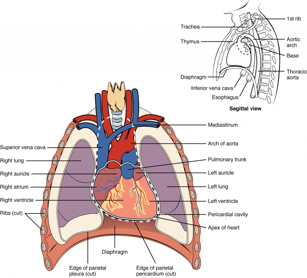
Information. The major cavities of the human body are the spaces left over when internal organs are removed. There are additional body cavities which we will only discuss in lecture. These are the cavities created by serous membranes-the pleural cavities, the pericardial cavity, and the peritoneal cavity-and the mediastinum.
1.04 Anatomical Terminology Body Cavities
The major organs of the abdomen include the small intestine, large intestine, and stomach. Together, these three turn nutrients into usable energy, as well as help dispose of solid waste. Major.
Cavities of body and anatomical compartment medical division outline
Body Membranes. The body cavities are lined with thin sheets of tissue called membranes, which cover and protect the various organs. The dorsal body cavity is lined with three layers of protective membranes (the dura mater, arachnoid, and pia mater), which are called the "meninges.". In 2014, I remember reading a news story about a nursing.

Dorsal Body Cavity Diagram Diagram Media
Thoracic wall The first step in understanding thorax anatomy is to find out its boundaries. The thoracic, or chest wall, consists of a skeletal framework, fascia, muscles, and neurovasculature - all connected together to form a strong and protective yet flexible cage.. The thorax has two major openings: the superior thoracic aperture found superiorly and the inferior thoracic aperture.
Pictures Of Cavity, Anatomical
Abdominal Body Cavity: Labeled diagram of the abdominal cavity showing the small intestine (brown) covered by the visceral peritoneum (inner green layer), and the parietal peritoneum (outer red layer) lining the abdominal cavity. The peritoneal cavity is the potential space between the visceral and parietal peritoneum and contains peritoneal fluid.
Dorsal Cavity Definition, Organs and Function Biology Dictionary
The abdomen is the body region found between the thorax and the pelvis. Its superior aperture faces towards the thorax, enclosed by the diaphragm. Inferiorly the abdomen is open to the pelvis, communicating through the superior pelvic aperture (pelvic inlet). These two apertures, together with abdominal walls, bound the abdominal cavity.
Major Cavities Of The Body cloudshareinfo
The abdominal cavity is where the majority of the body's organs lie. These are sometimes referred to as the "viscera", and they include organs like the liver, stomach, spleen, pancreas, kidneys and others involved in digestion, metabolism, and filtering of the blood. A special membrane holds all of these organs in place and is called the.
