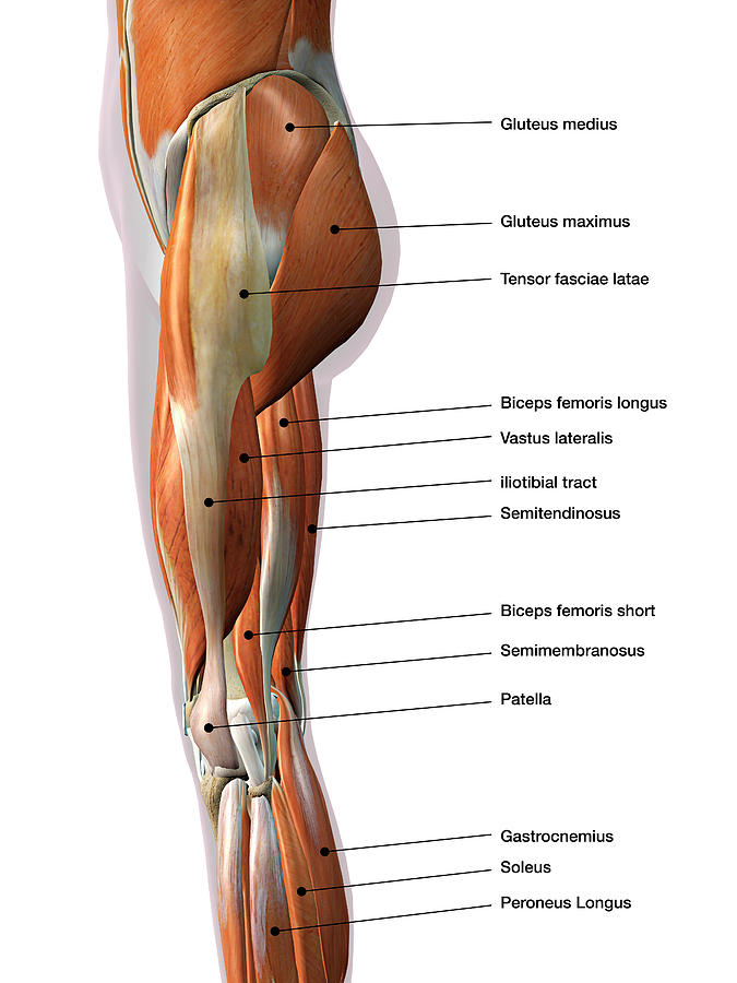
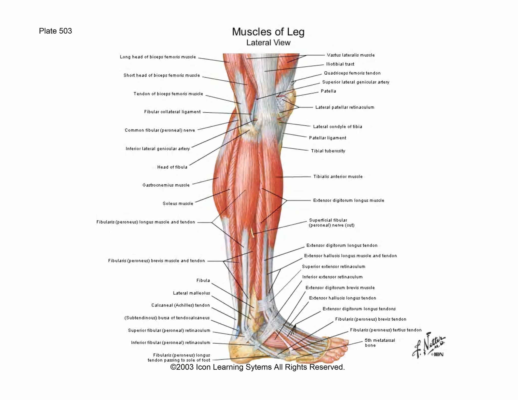
A List Of All The Muscle Names In The Legs Lateral view of a pair of
Leg muscles (Musculi cruris) Anatomically, the leg is defined as the region of the lower limb below the knee. It consists of a posterior, anterior and lateral compartment. In accordance, the muscles of the leg are organized into three groups: Anterior (dorsiflexor) group, which contains the tibialis anterior, extensor digitorum longus.

Leg muscles pain
1) Tibialis anterior (shin splints muscle) a. Actions: dorsiflexion, inverts foot (supports medial longitudinal arch) b. Innervation: Deep peroneal nerve. c. Origin: from lateral condyle and upper tibia. d. Insertion: to medial cuniform bone (tarsal) and 1st metatarsal bone. 2) Extensor digitorum longus.
Muscular System Anatomy and Physiology Nurseslabs
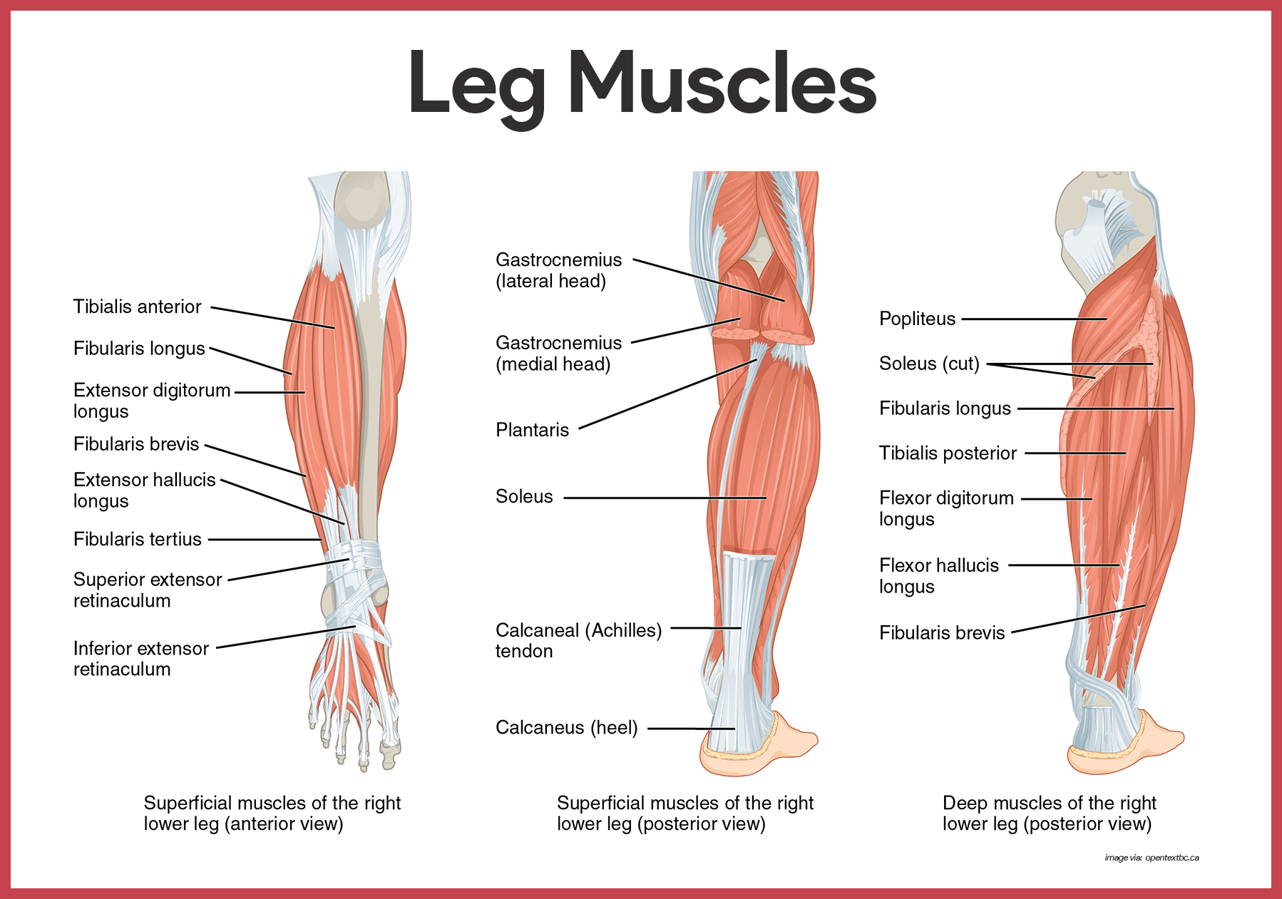
The posterior muscles are natural antagonists to the anterior muscle group. Generally, their main functions are plantarflexion, inversion of the foot, and flexion of the toes. Additionally, the natural tension of these muscles, especially the tibialis anterior, supports the medial arch of the foot. Namely, the deep flexor muscles of the leg are.

Image result for lower leg muscles lateral view Muscle anatomy, Leg
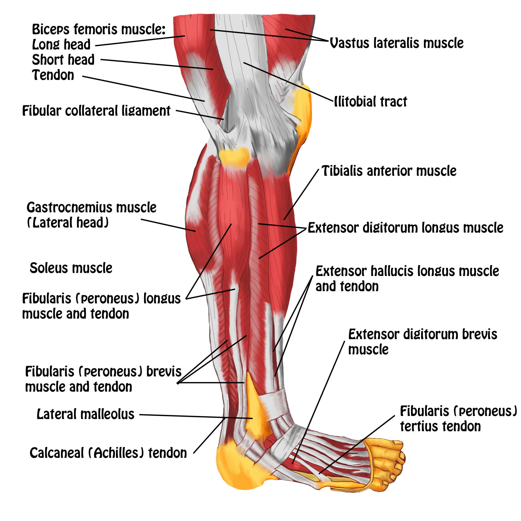
Gastrocnemius (calf muscle): One of the large muscles of the leg, it connects to the heel. It flexes and extends the foot, ankle, and knee. Soleus: This muscle extends from the back of the knee to.

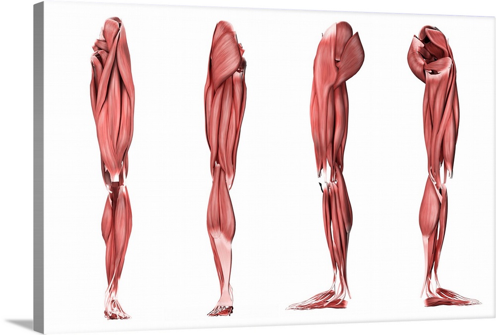
Leg muscle anatomical structure, labeled front, side and back view
Located inferior to the knee are a number of muscles that move the ankle, foot, and toes. The calf muscles, including the gastrocnemius and soleus, join to form the strong calcaneal (Achilles) tendon of the heel and attach to the calcaneus bone in the heel. These muscles contract to plantar flex the foot — such as when standing on your.
Lateral view of a pair of legs
The impact of foot position on electromyographical activity of the superficial quadriceps muscles during leg extension. Journal of strength and conditioning research, 19(4), 931-938.

leg muscle and tendon diagram Google Search MUSCLES AND ANATOMY
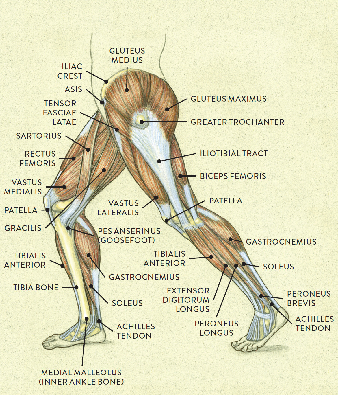
The knee joins the upper leg and the lower leg. It's also the largest joint in the body. In addition to bearing the weight of the upper body, the knee allows for walking, running, and jumping.

BIO201Leg Muscles Leg muscles anatomy, Muscle anatomy, Anatomy
The posterior compartment of the leg contains seven muscles and can be subdivided into superficial and deep compartments.. The muscles in this compartment act to plantarflex and invert the foot. They are innervated by the tibial nerve (a branch of the sciatic nerve). Blood supply chiefly from the posterior tibial artery. In this article, we shall look at the anatomy of the muscles in the.
Names Of Human Muscles With Illustration Human leg muscles
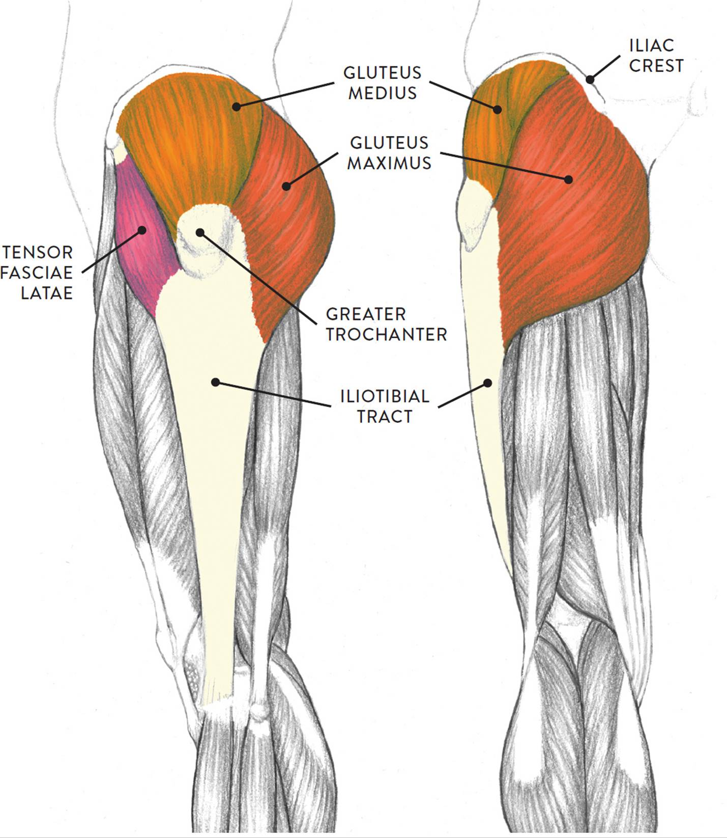
Side Leg Muscles. Your inner leg muscles or inner thigh muscles are known as your adductor muscles, which include the pectineus, adductor longus, adductor brevis, and gracilis. This group of leg muscles is responsible for bringing your thigh toward the center of your body, as well as rotating the thigh bone. Kailey Whitman.
Muscles of the Leg and Foot Classic Human Anatomy in Motion The
This cord-like muscle runs the length of your inner thigh from your pelvis to the inside of your knee. The adductors work virtually any time your legs are active, whether for standing, squatting, lunging, and most other leg moves. You can work them directly with inner-thigh leg raises and Swiss Ball squeezes.

Leg Muscle Diagram Side View / Pin on foot pain 3d human upper leg
One side of the obliques contracting can create lateral flexion. It also helps in pulling in the abdomen. The two muscles on either side of the chest come together to form a fibrous sheet. These muscles help the rectus abdominis to keep the abdominal organs in place. Gastrocnemius. The large muscle of the posterior part of the lower leg.
Å! 46+ Grunner til Deigram Of Outside Leg Muscles Start with a wide
The muscles in your upper and lower legs work together to help you move, support your body's weight and allow you to have good posture. They enable you to do big movements, like running and jumping. They also help you with small movements, like wiggling your toes. Leg muscle strains are common, especially in the hamstrings, quads and groin.

Leg Muscle Anatomical Structure, Labeled Front, Side And Back View
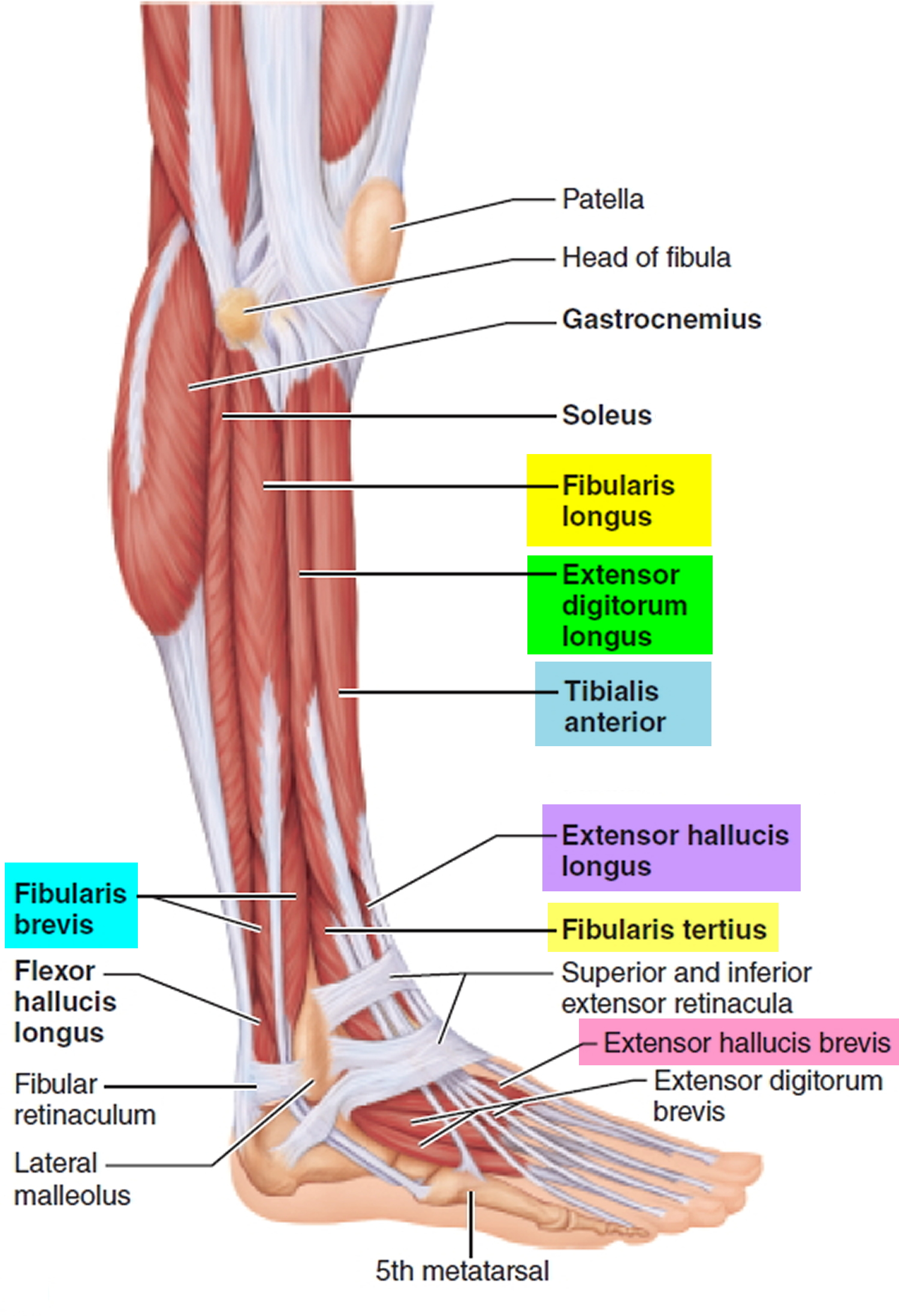
Anatomy of the Lower Leg. Your muscles in the lower leg are supported by two very strong, long bones: the fibula and the tibia (shinbone). The tibia is stronger and more prominent than the fibula. It is located toward the middle of the lower leg. The fibula, or calf bone, is smaller and located on the lower leg's outside.
Foot Drop Causes, Foot Drop Nerve, Symptoms, Prevention & Treatment
Muscles in the Lateral Compartment of the Leg. View Article. Muscles in the Posterior Compartment of the Leg. View Article. Anatomy Video Lectures. START NOW FOR FREE. TeachMe Anatomy. Part of the TeachMe Series.
Upper Leg Muscles And Tendons Tennis Leg and Achilles Tendonitis
Learn about different leg muscles and tendons and leg muscle anatomy: leg extensors, posterior leg muscles, extrinsic foot muscles, and intrinsic foot muscles. Updated: 11/21/2023 Table of Contents
Leg Muscle Diagram Side Beinschmerzen Symptome, Vorbeugung und
Lower extremity (anterior view). The posterior tibial artery gives off a crucial branch called the fibular/peroneal artery which mainly supplies the muscles of the leg. The tibial arteries originate from the popliteal artery.. but still on the plantar side of the foot, we meet the muscles of the lateral compartment: abductor digiti minimi
